Author Archives: Floyd Miracle

SVT with Aberrancy or Ventricular Tachycardia?

Differentiating between SVT with aberrancy and ventricular tachycardia (VT) can be a challenging task, even for experienced healthcare providers. This blog aims to clarify the distinction between these two cardiac arrhythmias, emphasizing the potential dangers of misdiagnosis and the importance of accurate identification. By understanding the concepts of aberrant conduction and the characteristics of SVT and VT, healthcare professionals can improve patient care and outcomes.

What Is SVT With Aberrancy?

The term “SVT with aberrancy” tends to throw many providers off, so let’s start by defining SVT using the 2015 ACC/AHA/HRS Guidelines as a reference.

“An umbrella term used to describe tachycardias (atrial and/or ventricular rates in excess of 100 bpm at rest), the mechanism of which involves tissue from the His bundle or above. These SVTs include inappropriate sinus tachycardia, AT (including focal and multifocal AT), macroreentrant AT (including typical atrial flutter), junctional tachycardia, AVNRT, and various forms of accessory pathway-mediated reentrant tachycardias. In this guideline, the term does not include AF.”

This is important because many of us were taught a narrow, complex rhythm: “must be SVT if the rate is over 150,” which can lead to inappropriate therapies. In reality, sinus tachycardia is a form of SVT, and the rate can easily exceed 150. A good rule of thumb to estimate the maximum sinus rate is 220 minus age, but that can vary by 10-15%, which is a lot.

What most people really mean when they call a rhythm “SVT” is AV Nodal Reentrant Tachycardia or AVNRT, which is a reentrant rhythm in or around the AV node. This arrhythmia is usually stable, and the prognosis is much more favorable than that of VT. It is usually treated with vagal maneuvers or adenosine.

What Does Aberrancy Mean?

You can think of “aberrancy” as abnormal conduction. When something is aberrant, it “departs from the right, normal, or usual course.”

Because the right bundle branch tends to have a slightly longer refractory period than the left bundle branch, at higher rates the right bundle branch may not be fully recovered from the previous cardiac cycle, which results in a right bundle branch block pattern.

Even though right bundle branch block aberrancy is more common than left bundle branch block aberrancy, both are possible. Additionally, we know that many patients have underlying bundle branch block, including bifascicular block, at baseline.

When a patient with a bundle branch block experiences SVT, the result is a wide complex tachycardia.

Is Aberrant Conduction Dangerous?

While aberrant conduction itself is not inherently dangerous, the potential for misdiagnosis is a significant concern. Mistaking SVT with aberrancy for VT can lead to the administration of antiarrhythmic medications or cardioversion, which can be harmful if the underlying rhythm is SVT.

SVT With Aberrancy vs. VT

Differentiating between SVT with aberrancy and VT can help determine the appropriate treatment. Several factors can help distinguish between the two, including the patient’s clinical presentation, the appearance of the ECG, and the response to specific medications. While there are diagnostic criteria to help rule in VT, there are currently no definitive criteria to safely rule out VT.

It is important to approach wide complex tachycardias with caution and to consider the possibility of both SVT with aberrancy and VT. Obtaining a 12-lead ECG before and after treatment can aid in diagnosis. In cases of unstable wide complex tachycardia, immediate synchronized cardioversion is often necessary.

Can You Differentiate Between SVT With Aberrant Conduction and VT?

The short answer is yes, but it can be very difficult, and even experienced clinicians can misdiagnose VT as SVT with aberrancy!

This can lead to clinical misadventure. In particular, treating a wide complex tachycardia with a calcium channel blocker is a dangerous decision that could have fatal consequences for your patient.

There are good criteria to help rule-in, or tip the scales in favor of VT, but none to safely rule-out VT.

See also: Myths and Cognitive Biases in Interpretation of Wide Complex Tachycardias

Consider the following case:

EMS is dispatched to an 83-year-old female who contacts 9-1-1 after she wakes up with a “racing heart” and shortness of breath.

Past medical history includes myocardial infarction and hypertension.

On initial assessment the patient is found to be alert and oriented to person, place, time, and event. The skin is pale but warm and dry. Radial pulses are very rapid but surprisingly strong. Breath sounds are clear bilaterally.

She is placed on the cardiac monitor, and the following rhythm strip is obtained.

regular complex tachycardia at a rate of ~ 230 bpm

Figure 1: There is a wide and regular complex tachycardia at a rate of ~ 230 bpm.

The patient is placed on oxygen via nasal cannula, and IV access is established while vital signs are obtained.

  • RR: 24
  • HR: Too fast to count
  • NIBP: 112/72
  • SpO2: 97%
  • Temp: 98.3 F / 36.8 C

Why should you presume that this rhythm is ventricular tachycardia?

  • VT accounts for 80% of all cases of WCT
  • If the patient has a previous cardiac history, the predictive value can go up over 90%
  • An age greater than 35 years has a sensitivity of 92%

Treatment

A 12 lead ECG is obtained.

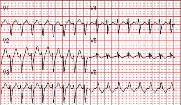
Figure 2: There is a regular wide complex tachycardia at a rate of about 230 without sinus P waves. There is a LBBB pattern in lead V1. However, we would not consider this to be a “typical” LBBB pattern due to the normal axis in the frontal plane and the presence of a small S-wave in lead I.

Amiodarone 150 mg is given over 10 minutes.
A rhythm change is noted, and the following 12-lead ECG is obtained.

Figure 3: Now there is sinus tachycardia with virtually identical QRS morphology.

Once the patient converts to sinus tachycardia (and after a sigh of relief), paramedics compare the two 12-lead ECGs. The axis and QRS morphology are noted to be exactly the same.
The diagnosis? SVT with aberrancy!

It is safe to conclude that this patient had a conduction defect at baseline, which is what caused the complexes to be wide during the tachycardia.

Retrospectively, adenosine would have been safe and likely effective. In many cases, it can be considered as a first line therapy for undifferentiated wide complex tachycardia, and may have some diagnostic utility when considered in the context of other findings.

Take the Next Step in Cardiac Care With ACLS Medical Training

Accurately differentiating between SVT with aberrancy and ventricular tachycardia is crucial for effective and safe patient care. Misdiagnosis can have severe consequences. By understanding the key characteristics of each arrhythmia and utilizing appropriate diagnostic tools, healthcare providers can improve their ability to recognize and manage these complex cardiac conditions.

To further enhance your knowledge and skills in advanced cardiac life support, consider enrolling in our online ACLS certification course. By investing in your ACLS certification, you’ll be better equipped to handle a wide range of cardiac emergencies and provide optimal care for your patients. Head to our website to get started today!

References

Alzand BCrijns H. Diagnostic criteria of broad QRS complex tachycardia: decades of evolution. Europace. 2010;13(4):465-472

Neumar R, Otto C, Link M et al. Part 8: Adult Advanced Cardiovascular Life Support: 2010 American Heart Association Guidelines for Cardiopulmonary Resuscitation and Emergency Cardiovascular Care. Circulation. 2010;122(18_suppl_3):S729-S767
CPR First? Or Defibrillation First?

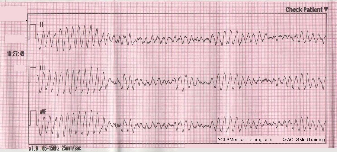
Ventricular Fibrillation is considered the most favorable cardiac arrest rhythm, and if treated promptly can result in ROSC with a favorable neurological outcome. Most survival rates are reported using witnessed arrest with a shockable rhythm as opposed to asystole or PEA, as the outcomes of these rhythms are comparatively very poor.

The Resuscitation Academy mantra “everyone in VF survives” has been adopted by many EMS systems around the world to emphasize that these patients can and do survive, and it’s up to us to save them.

Major advances have been made over the past 10 years but CPR and defibrillation are still the bedrock of resuscitation science. The attributes of high-quality CPR were re-affirmed in the 2015 AHA ECC Guidelines.

  • Ensuring adequate rate (100-120)
  • Ensuring adequate depth (2 to 2.4” or 5 to 6 cm)
  • Allowing full chest recoil (avoid leaning)
  • Minimizing interruptions to chest compressions
  • Avoiding excessive ventilations

Is CPR Before Defibrillation Dogmatic?

In the context of a witnessed arrest by a trained first responder or bystander who has an AED or manual defibrillator, the importance of early defibrillation is irrefutable. We have been told repeatedly that early defibrillation saves lives.

I initially began my research under the assumption that providing 1.5 to 3 minutes of CPR before defibrillation provides oxygen and nutrients to the heart therefore making defibrillation more likely to be successful. However, recent evidence suggests that performing chest compressions while setting up the defibrillator and charging the capacitor may be adequate.

A “CPR first” approach is rooted in evidence suggesting the existence of 3 time-sensitive phases of VF arrest.

  1. Electrical phase (0-4 minutes)
  2. Circulatory phase (5-10 minutes)
  3. Metabolic Phase (> 10 minutes)

Researchers suggested that a period of CPR prior to defibrillation might confer a benefit during the so-called “circulatory phase” of the cardiac arrest.

Evolution of American Heart Association Recommendations

Because it is rare for EMS to arrive on scene during the electrical phase, the 2005 AHA ECC Guidelines made this recommendation:

When an out-of-hospital cardiac arrest is not witnessed by EMS personnel, they may give about 5 cycles of CPR before checking the ECG rhythm and attempting defibrillation (Class IIb). One cycle of CPR consists of 30 compressions and 2 breaths. When compressions are delivered at a rate of about 100 per minute, 5 cycles of CPR should take roughly 2 minutes (range: about 1½ to 3 minutes). This recommendation regarding CPR prior to attempted defibrillation is supported by 2 clinical studies (LOE 2, LOE 3) of adult out-of-hospital VF SCA. In those studies when EMS call-to-arrival intervals were 4 to 5 minutes or longer, victims who received 1½ to 3 minutes of CPR before defibrillation showed an increased rate of initial resuscitation, survival to hospital discharge, and 1-year survival when compared with those who received immediate defibrillation for VF SCA. One randomized study, however, found no benefit to CPR before defibrillation for non-paramedic-witnessed SCA.

Fast forward 10 years to the 2015 Guidelines.

Observational clinical studies and mechanistic studies in animal models suggest that CPR under conditions of prolonged untreated VF might help restore metabolic conditions of the heart favorable to defibrillation…others have suggested that prolonged VF is energetically detrimental to the ischemic heart, justifying rapid defibrillation attempts regardless of the duration of arrest.

Evidence summary

Five RCTs, 4 observational cohort studies, 3 meta-analyses, and 1 subgroup analysis of an RCT addressed the question of CPR before defibrillation. The duration of CPR before defibrillation ranged from 90 to 180 seconds, with the control group having a shorter CPR interval lasting only as long as the time required for defibrillator deployment, pad placement, initial rhythm analysis, and AED charging. These studies showed that outcomes were not different when CPR was provided for a period of up to 180 seconds before attempted defibrillation compared with rhythm analysis and attempted defibrillation first for the various outcomes examined, ranging from 1-year survival with favorable neurologic outcome to ROSC. Subgroup analysis suggested potential benefit from CPR before defibrillation in patients with prolonged EMS response intervals (4 to 5 minutes or longer) and in EMS agencies with high baseline survival to hospital discharge, but these findings conflict with other subset analyses.  Accordingly, the current evidence suggests that for unmonitored patients with cardiac arrest outside of the hospital and an initial rhythm of VF or pVT, there is no benefit from a period of CPR of 90 to 180 seconds before attempted defibrillation.

Specifically, the ROC PRIMED trial concluded that:

Among patients who had an out-of-hospital cardiac arrest, we found no difference in the outcomes with a brief period, as compared with a longer period, of EMS-administered CPR before the first analysis of cardiac rhythm.

The ROC Investigators subsequently found that EMS systems with a VF survival rate < 20% appeared to do better with an “analyze first” strategy. Conversely, EMS systems with a VF survival rate > 20% appeared to do better with a “analyze late” strategy.

Can the VF Waveform Determine the Likelihood of Successful Defibrillation?

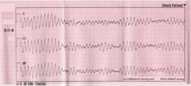
Ventricular fibrillation sometimes begins as ventricular tachycardia, and if left untreated deteriorates into fine VF. Fine VF predictably results in conversion to asystole or continued VF, but rarely to a perfusing rhythm.

Berg et al. performed a randomized, controlled trial using animals. After inducing VF in swine for 8 minutes, they were randomly assigned to either immediate defibrillation, or defibrillation provided after 90 seconds of CPR. Nine out of 15 attained ROSC in the CPR first group, and zero out of 15 who were defibrillated first resulted in ROSC. Their conclusion?

Pre-countershock CPR can result in substantial physiologic benefits and superior response to initial defibrillation attempts compared with immediate defibrillation in the setting of prolonged ventricular fibrillation.

Additionally, they determined there was a mathematical relationship between the VF waveform and chances of successful defibrillation. The animals who received CPR first had a much higher median frequency, and a much higher rate of ROSC than those that did not.

In the field, whether or not VF is “fine” or “coarse” is typically based on visual inspection of the waveform. What if there was a way to accurately determine which patients would benefit from defibrillation and those that would not, thus eliminating unnecessary pauses and ineffective shocks?

Callaway et al. and Eftestol et al. supported the theory that VF frequency and amplitude could be used to determine which patients will respond to countershock.

Eftestol et al. concluded:

CPR done by professionals can improve the chance for ROSC and ultimate survival of patients with prolonged cardiac arrest and significantly deteriorated myocardium. This study also indicates that CPR periods of 3 minutes might be better for the myocardium than shorter periods. Finally, together with the studies showing rapid deterioration of the myocardium in even a few seconds without CPR after a cardiac arrest, it gives the important message that periods without CPR (for ECG analysis, defibrillation charging, pulse checks, intubation attempts, etc) should be kept to a minimum. This is frequently not the case clinically.

As promising as this may have seemed, an article in Circulation by Freese et al. evaluated the theory of defibrillation based on waveform analysis, and the results were disappointing.

Use of a waveform analysis algorithm to guide the initial treatment of out-of-hospital cardiac arrest patients presenting in VF did not improve overall survival compared with a standard shock-first protocol. Further study is recommended to examine the role of waveform analysis for the guided management of VF.

The Bottom Line

The totality of the evidence suggests that defibrillation as soon as practicable (with the caveat that high quality chest compressions are performed while setting up the defibrillator) is equivalent to a prescribed interval of CPR prior to the first shock in most instances.

EMS systems that measure the “patient’s side to first shock” interval know that it usually takes at least 1 minute to power on the defibrillator, extend the cables, attach the pads, charge the capacitor, and deliver the shock. During that interval, there’s no reason that the patient can’t receive continuous chest compressions.

One benefit to emphasizing a “shock as soon as possible” approach is that it’s the same for EMS-witnessed cardiac arrest.

Alternatively, defibrillation can be delivered after the first 2-minute cycle. It seems likely that CPR quality plays a more important role than the exact timing of the first shock.

References

Baker PW, Conway J, Cotton C, Ashby DT, Smyth J, Woodman RJ, Grantham H; Clinical Investigators. Defibrillation or cardiopulmonary resuscitation first for patients with out-of-hospital cardiac arrests found by paramedics to be in ventricular fibrillation? A randomised control trial. Resuscitation. 2008;79:424–431. doi: 10.1016/j.resuscitation.2008.07.017.

Bradley SM, Gabriel EE, Aufderheide TP, Barnes R, Christenson J, Davis DP, Stiell IG, Nichol G; Resuscitation Outcomes Consortium Investigators. Survival increases with CPR by Emergency Medical Services before defibrillation of out-of-hospital ventricular fibrillation or ventricular tachycardia: observations from the Resuscitation Outcomes Consortium. Resuscitation. 2010;81:155–162. doi: 10.1016/j. resuscitation.2009.10.026.

Cobb LA, Fahrenbruch CE, Walsh TR, Copass MK, Olsufka M, Breskin M, Hallstrom AP. Influence of cardiopulmonary resuscitation prior to defibrillation in patients with out-of-hospital ventricular fibrillation. JAMA. 1999;281:1182–1188.

Freese J, Jorgenson D, Liu P et al. Waveform Analysis-Guided Treatment Versus a Standard Shock-First Protocol for the Treatment of Out-of-Hospital Cardiac Arrest Presenting in Ventricular Fibrillation: Results of an International Randomized, Controlled Trial. Circulation. 2013;128(9):995-1002. doi:10.1161/circulationaha.113.003273.

Gilmore C, Rea T, Becker L, Eisenberg M. Three-Phase Model of Cardiac Arrest: Time-Dependent Benefit of Bystander Cardiopulmonary Resuscitation. The American Journal of Cardiology. 2006;98(4):497-499. doi:10.1016/j.amjcard.2006.02.055.

Hayakawa M, Gando S, Okamoto H, Asai Y, Uegaki S, Makise H. Shortening of cardiopulmonary resuscitation time before the defibrilla- tion worsens the outcome in out-of-hospital VF patients. Am J Emerg Med. 2009;27:470–474. doi: 10.1016/j.ajem.2008.

Huang Y, He Q, Yang LJ, Liu GJ, Jones A. Cardiopulmonary resuscitation (CPR) plus delayed defibrillation versus immediate defibrillation for out-of-hospital cardiac arrest. Cochrane Database Syst Rev. 2014;9:CD009803. doi: 10.1002/14651858.CD009803.pub2.

Jacobs IG, Finn JC, Oxer HF, Jelinek GA. CPR before defibrillation in out-of-hospital cardiac arrest: a randomized trial. Emerg Med Australas. 2005;17:39–45. doi: 10.1111/j.1742-6723.2005.00694.x.

Kleinman M, Brennan E, Goldberger Z et al. Part 5: Adult Basic Life Support and Cardiopulmonary Resuscitation Quality. Circulation. 2015;132(18 suppl 2):S414-S435. doi:10.1161/cir.0000000000000259.

Koike S, Tanabe S, Ogawa T, Akahane M, Yasunaga H, Horiguchi H, Matsumoto S, Imamura T. Immediate defibrillation or defibrillation after cardiopulmonary resuscitation. Prehosp Emerg Care. 2011;15:393–400. doi: 10.3109/10903127.2011.569848.

Ma MH, Chiang WC, Ko PC, Yang CW, Wang HC, Chen SY, Chang WT, Huang CH, Chou HC, Lai MS, Chien KL, Lee BC, Hwang CH, Wang YC, Hsiung GH, Hsiao YW, Chang AM, Chen WJ, Chen SC. A randomized trial of compression first or analyze first strategies in patients with out-of-hospital cardiac arrest: results from an Asian community. Resuscitation. 2012;83:806–812. doi: 10.1016/j.resuscitation.2012.01.009.

Meier P, Baker P, Jost D, Jacobs I, Henzi B, Knapp G, Sasson C. Chest compressions before defibrillation for out-of-hospital cardiac arrest: a meta-analysis of randomized controlled clinical trials. BMC Med. 2010;8:52. doi: 10.1186/1741-7015-8-52.

Naloxone

Is there an Irrational Fear of Naloxone?

Naloxone

In the U.S. there has been a 286% increase in heroin-related overdose deaths since 2002. In 2014, a total of 47,055 drug overdose deaths occurred. Our EMS systems and emergency departments have felt the surge, along with a corresponding increase in the use of the drug naloxone.

Community officials have taken the initiative to reduce the number of overdose deaths by making naloxone available through non-traditional means. However, this has not been particularly well received by members of the medical community, particularly pre-hospital providers.

Discussion of the proper use of naloxone often sparks a passionate, sometimes contentious debate, usually focused around the deleterious side effects and inappropriate use of naloxone.

Hopefully, a review of the literature will make providers less apprehensive about the expanded use of naloxone and spark a healthy debate of the current standard of care.

Treatment of an opioid overdose

A common strategy for treating an opioid-related overdose is to initiate BVM ventilation to treat respiratory depression and then titrating naloxone until the patient’s respiratory effort becomes adequate but not to the point of the patient becoming lucid.

Should patients be given enough naloxone to regain consciousness?

There was a time when I passionately advocated for allowing patients to remain somnolent in the setting of a suspected opioid overdose, but I changed my mind after encountering this emergency more frequently.

Like many things in medicine, it sounds good in theory, but it doesn’t translate easily into practice. Almost every attempt I made at titrating naloxone until the respiratory depression improved resulted in the patient becoming completely lucid.

What about adverse outcomes associated with naloxone administration?

Many emergency providers are apprehensive about giving naloxone due to undesirable side effects. Patients regaining consciousness after naloxone administration may experience many symptoms that are collectively referred to as Acute Withdrawal Syndrome (AWS).

Patients with AWS may exhibit nausea, vomiting, tachycardia, diarrhea, hypertension, nervousness, and restlessness. The degree of withdrawal symptoms is relatively proportional to the amount of naloxone given, so smaller amounts will normally result in less serious withdrawal symptoms.

Rare but serious complications like cardiac arrest, seizures, acute pulmonary edema, and violent behavior are sometimes offered as reasons for not using naloxone (or not giving enough for the patient to regain consciousness). However, it is likely that these complications have been overstated, as the evidence has not been reproducible.

Osterwalder (1996) investigated subjects treated with naloxone from 1991-1993. Six out of 453 patients experienced severe adverse effects. One suffered asystole, three generalized convulsions, one pulmonary edema, and one violent behavior.

However, Burris (2000) reported in the International Journal of Drug Policy that “more recent research suggests that complications are exceedingly rare, that past reports of complications may have been erroneous, or that complications occur, if at all, in patients with pre-existing heart disease”.

Yearly et al. (1990) conducted a retrospective study of over 800 prehospital records of patients who received initial IV doses of 0.4-0.8 mg of naloxone and found that no patients experienced ventricular tachycardia, fibrillation, or asystole. There was one generalized tonic-clonic seizure in a patient with a history of seizures. The authors concluded that smaller doses of naloxone are not warranted.

What if a patient refuses transport after regaining consciousness?

Typically patients are transported to the hospital after regaining consciousness following the administration of naloxone. However some patients refuse, against the advice of treating paramedics.

One might expect there to be a high mortality for patients who refuse transport, since it’s widely known that the half-life of naloxone is shorter than the half-life of the opioid. However, current data suggest that a patient can refuse transport without serious consequences.

In San Antonio, Wampler et al. (2011) conducted a review of 595 patients treated with naloxone in a large fire-based EMS system who refused transport to the hospital. The San Antonio protocol consisted of giving naloxone, 2 mg IM, 2 mg IV, and an additional 2mg IM with patient consent. None were found in the Medical Examiner’s Office database two-days after refusing transport. Although 9 of the patients subsequently died, the shortest time interval was four days after treatment.

Vike et al (2003) conducted another retrospective review comparing prehospital and medical examiner databases. In the prehospital database there were 998 patients who had received naloxone and refused transport. In the medical examiner’s database there were 601 recorded opioid overdose deaths. None of them had been treated with naloxone within 12 hours of death.

Conclusion

  • Naloxone is a safe and effective treatment for opioid overdose.
  • Expanded use of naloxone is unlikely to cause an increase in adverse outcomes.
  • Criteria should be established to help predict patients that can safely refuse transport to the hospital.

References

Burris S, Norland J, Edlin B. Legal aspects of providing naloxone to heroin users in the United States. International Journal of Drug Policy. 2001;12(3):237-248.

Kim D, Irwin K, Khoshnood K. Expanded Access to Naloxone: Options for Critical Response to the Epidemic of Opioid Overdose Mortality. Am J Public Health. 2009;99(3):402-407.

Opioid OD patients revived with naloxone who refuse further treatment do not die. EMSWorld.com. 2016. Available at: http://www.emsworld.com/article/10284002/opioid-od-patients-revived-with-naloxone-who-refuse-further-treatment-do-not-die. Accessed July 12, 2016.

Wermeling D. Review of naloxone safety for opioid overdose: practical considerations for new technology and expanded public access. Therapeutic Advances in Drug Safety. 2015;6(1):20-31.

Wampler D, Molina D, McManus J, Laws P, Manifold C. No Deaths Associated with Patient Refusal of Transport After Naloxone-Reversed Opioid Overdose. Prehospital Emergency Care. 2011;15(3):320-324.

Boyer E. Management of Opioid Analgesic Overdose. New England Journal of Medicine. 2012;367(2):146-155.

Vilke G, Buchanan J, Dunford J, Chan T. Are heroin overdose deaths related to patient release after prehospital treatment with naloxone?. Prehospital Emergency Care. 1999;3(3):183-186.

How to Master BVM Ventilation

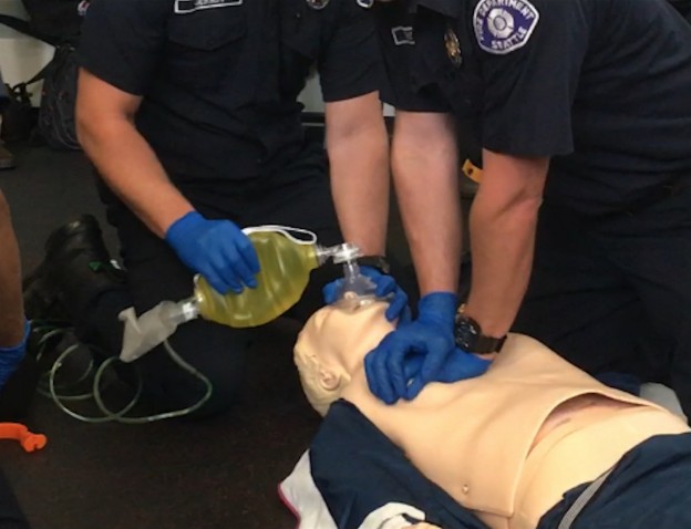
Bag mask ventilation is the cornerstone of airway management.

It’s often considered a basic procedure, but there is nothing “basic” about BVM ventilation. Skill acquisition requires extensive training and experience. It’s not pretty, sexy, or glamorous. Most people perform it poorly even though it’s an essential part of good airway management.

We often relegate the skill to a new or junior provider, and when the saturation drops we attribute it to the patients’ acuity and not to failure to provide adequate oxygenation and ventilation.

The AHA recognizes that bag mask ventilation “is a challenging skill that requires considerable practice for competency.”

When performed in an emergency, respiratory failure or arrest is often imminent. Because it is a BLS skill we toss a BVM to our partner while we prepare our intubation equipment. The mask is placed on the patient’s face and ventilations are administered too aggressively or ineffectively.

When this is not recognized the airway may become flooded with gastric contents during the intubation attempt making more difficult if not impossible. Aspiration occurs, hypoxia worsens, and the patient is at higher risk of experiencing cardiac arrest.

It is not widely appreciated that BVM ventilation is often ineffective. One assumes that it works better than it actually does without appreciate education and training, which is often lacking.

Early in my career I would place the mask on the patient’s face and apply the CE technique without any objective measurement of how well it was working.

How do you Know When Ventilations are Effective?

Clinical detection of adequate ventilation is notoriously difficult. So what is the litmus test for gas exchange at the alveolar level? ETCO2 of course!

The ETCO2 sensor fits perfectly between the bag and mask.

In Emergency Medicine we want the technique that is most likely to be successful the first time. The traditional CE method is not always the best technique. Some will be quick to contest that assertion, and a few years ago I would have agreed with you.

Then I watched a video on EmCrit made by Reuben Strayer.

There are three main factors that contribute to poor BVM ventilation.

  • Poor mask seal
  • Improper positioning
  • Excessive rate and volume

Poor Mask Seal

When using the traditional CE technique, pressure is not distributed equally across the mask. This means that when using your left hand, there is a tendency for air to leak between the mask and the right side of the patient’s mouth, which often goes unrecognized.

Improper Positioning

Because of the inherit difficulty maintaining a quality seal, and because maintaining a seal is fatiguing, the tendency is to push the mask onto the face. The mouth is then closed shut, leaving the nares as the only route of ventilation. Obstructive soft tissues of the pharynx collapse, blocking the glottic opening.

A superior technique was introduced 11 years ago in the 2005 AHA Guidelines.

“Bag-mask ventilation is most effective when provided by 2 trained and experienced rescuers. One rescuer opens the airway and seals the mask to the face while the other squeezes the bag. Both rescuers watch for visible chest rise.”

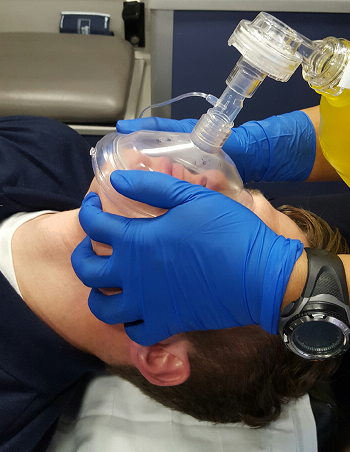
The two handed technique is sometimes referred to as the thenar eminence (TE), or “two thumbs down” technique.

The fingers are used to bring the jaw to the mask, while the palms and thumbs maintain a mask seal. This offers a mechanical advantage to the CE technique and allows better recognition of air leaks.

Gerstein (2013) compared the effectiveness of the CE and TE technique when performed by novice clinicians and found:

“The TE facemask ventilation grip results in improved ventilation over the EC grip in the hands of novice providers.”

A few weeks ago I attended a cadaver lab in Baltimore. The first skill we practiced was BVM ventilation. Our group leader has us try the CE technique first, then TE. The chest was open and lungs exposed so we could see the effectiveness of our ventilations.

Six people were in my group, and no one was able to inflate the lungs using the CE technique despite multiple attempts. However, the lungs were inflated every time, every attempt, for every person when using the TE technique!

Excessive Rate and Tidal Volume (Hyperventilation)

Even when trying to be cognizant of rate and tidal volume, there can be a huge difference in what you think you’re doing, and what you’re actually doing.

This was proven in the Milwaukee study, in which Paramedics were taught to ventilate at the appropriate rate during cardiac arrest. They retrospectively looked at the ventilation rates objectively and found the average rate was 30 breaths/min!

An excessive rate and tidal volume isn’t only deleterious for patients in cardiac arrest, but increases the likelihood of exceeding the pressure of the lower esophageal sphincter, delivering large tidal volumes of air to the stomach.

This also was mentioned back in the 2005 AHA Guidelines:

“Gastric inflation often develops when ventilation is provided without an advanced airway. It can cause regurgitation and aspiration, and by elevating the diaphragm, it can restrict lung movement and decrease respiratory compliance. Air delivered with each rescue breath can enter the stomach when pressure in the esophagus exceeds the lower esophageal sphincter opening pressure. Risk of gastric inflation is increased by high proximal airway pressure and the reduced opening pressure of the lower esophageal sphincter. High pressure can be created by a short inspiratory time, large tidal volume, high peak inspiratory pressure, incomplete airway opening, and decreased lung compliance.”

To prevent gastric inflation the airway must be kept open, and breaths delivered slowly…very slowly. Based on my observations no one delivers breaths slow enough. When your own heart rate is going 150 beats per minute, waiting 6 seconds to deliver a breath feels like forever! I often tell someone who is bagging to fast to deliver a breath every 10 seconds and even then they often ventilate too fast.

How do we slow down? Well, if the patient is intubated they could be placed on the ventilator. But since we’re talking about facemask ventilation, consider purchasing a timing light that goes on the end of the BVM, or use a metronome. You could also try counting, “one, one thousand…two, one thousand…three, one thousand…” and so on.

In addition to delivering breaths too fast, we deliver too much. The average volume of an adult BVM is 1600 milliliters! Squeezing the bag until opposite sides of the BVM touch isn’t necessary! It’s recommended that only 1/3 of the bag be compressed to give a large enough tidal volume. Any more and the pressure is too much for the rigid trachea to accommodate, and the esophagus is more than happy to accept the rest!

BVM Ventilation during Cardiac Arrest

If you’re doing 30:2 during BLS CPR you don’t have the luxury of providing breaths slowly. The goal should be to have compressions resumed within 3 seconds, and to do that the breaths can’t be given quickly or it will take 5 or 6 seconds!

The goal should be “little bag squeeze, little bag squeeze” with full release between squeezes. Intrathoracic pressure stays elevated without a full release, and we know that increased intrathoracic pressure impedes venous return.

Conclusion

  • BVM ventilation is a difficult skill for providers at all levels and specialties.
  • The traditional CE method is not very effective, and sometimes totally ineffective.
  • Use ETCO2 as an objective measurement.
  • Adopt the “two thumbs down” technique
  • Deliver breaths slowly
  • Only compress 1/3 of the bag
  • Give breaths quickly during cardiac arrest, but allow full release of BVM

References
“Beginner Facemask Ventilation Techniques | Emsworld.Com”. EMSWorld.com. N.p., 2016. Web. 17 Mar. 2016.
Gerstein NS, et al. “Efficacy Of Facemask Ventilation Techniques In Novice Providers. – Pubmed – NCBI”. Ncbi.nlm.nih.gov. N.p., 2016. Web. 17 Mar. 2016.
“Part 4: Adult Basic Life Support”. Circulation 112.24_suppl (2005): IV-19-IV-34. Web. 17 Mar. 2016

Cardiac Cath Lab Activation for Subtle Acute Inferior STEMI

EMS responds to a 78 year old male complaining of chest pain. On initial observation the patient is pale, cool, and diaphoretic. He says to the treating paramedic, “I think I’m having a heart attack.” He states that he was watching TV when he felt a crushing pain that radiated to his arm and jaw.

Pertinent Medical History

  • CABG X 2
  • Stents
  • CVA
  • Hernia Repair

Vital Signs

  • RR: 17
  • HR: 75
  • NIBP: 146/80
  • SpO2: 98% on room air

The following 12 Lead was acquired.

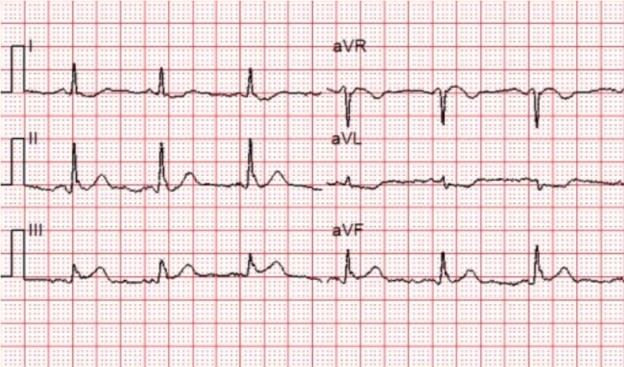
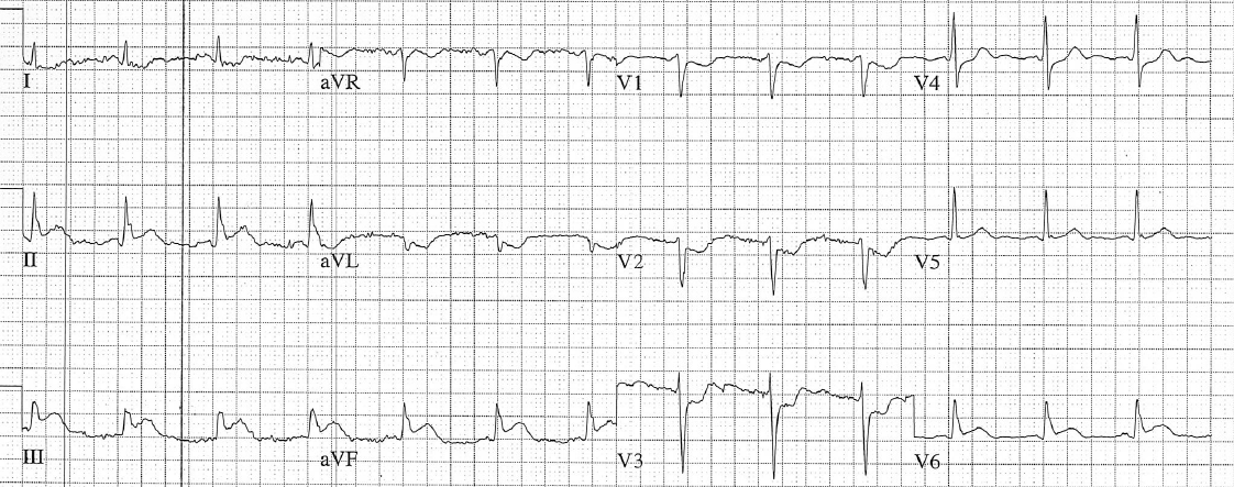
Sinus rhythm with ST depression in leads I, aVL, V2-V4, and 1mm of elevation in lead III.

Is this ECG diagnostic for an acute STEMI?

The Guidelines require new ST-segment elevation, measured at the J-point in at least 2 contiguous leads of ≥ 2 mm (men) or ≥ 1.5 mm (women) in leads V2-V3 and/or ≥ 1 mm in other contiguous leads or the limb leads.

Using this criteria, the EKG is only about 45% sensitive for an acute MI. That means that if we strictly went by mm criteria only 45 out of every 100 patients experiencing acute STEMI would be picked up on the 12-lead ECG. That’s a lot of missed occlusions.

Conversely, many patients have ST-segment elevation at baseline that is not the result of acute coronary occlusion.

In this case lead III is the only lead that if blown up may have about 1 mm of ST-segment elevation.

According to the treating paramedic, the patient presented with classic signs and symptoms. It may be important to note that this was an experienced paramedic and his gut told him the patient was experiencing a heart attack, so he activated the cardiac cath lab.

There were no observable changes on serial prehospital 12-lead ECGs. However, there was a difference noted on the 12-lead ECG obtained on arrival in the Emergency Department.

There is worsening of ST-segment elevation in the inferior leads, new ST-segment elevation in lead V6, and the reciprocal ST-segment depression in leads V1-V3 now looks diagnostic for posterior extension.

When I showed the initial ECG to other providers, most were quick to point out that the ECG was “not diagnostic” because of the absence of ST-segment elevation in 2 contiguous leads.

This is not surprising. Most ECG courses spend a lot of time going over “slam dunk” ECGs with significant ST-segment elevation so that students don’t learn to appreciate subtle signs of acute STEMI.

Looking at the first ECG we can be almost certain that the patient is experiencing acute inferior STEMI even though it does not meet millimeter criteria.

The ST-segment depression must be explained. One of the most salient points that impacted my understanding of subtle occlusions is that ischemia does not localize.

We have all been taught that subendocardial ischemia presents as ST-segment depression whereas transmural (epicardial) ischemia from coronary occlusion typically manifests as ST-segment elevation.

Subendocardial ischemia typically presents with ST-segment elevation in lead aVR with widespread ST-segment depression that does not “localize” to a particular set of leads.

In this case there is isolated ST-segment depression in the high lateral leads (I and aVL) and in the anterior leads (V1-V4).

This means that the most probable explanation for the ST-segment depression is not ischemia, but reciprocal changes from an inferior-posterior STEMI!

Always consider the possibility that a subtle STEMI is present when there is isolated ST-segment depression on the 12-lead ECG.

ST-segment depression in lead aVL is highly sensitive for acute inferior STEMI. If there is ST-segment depression in lead aVL you should consider the possibility of acute inferior STEMI, especially if there is ST-segment elevation in lead III.

ST-segment depression in lead aVL can precede ST-segment elevation in the inferior leads!

However, ST-segment depression in lead aVL can also be caused by so-called “secondary” ST/T-wave abnormalities. However, we can easily rule those out in this case because:

  • The QRS is not wide – There’s no bundle branch block or WPW pattern
  • There’s no high voltage – Left ventricular hypertrophy is not present
  • The QRS/T angle is not “wide” – Normally in the presence of a secondary ST/T-wave abnormality there is a general pattern of T-wave discordance. That means that when the majority of the QRS complex is positive the T-wave should be negative. When the majority of the QRS complex is negative, the T-wave should be positive.

Therefore the ST-segment changes in the initial ECG should be considered primary — due to acute STEMI.

Left ventricular hypertrophy is the most common of STEMI mimics, so it may be worthwhile to review the criteria here.

What about activating the cardiac cath lab for subtle STEMIs?

Cardiac cath lab activation should be reserved for clear-cut STEMI.

Most prehospital protocols require some combination of millimeter criteria, reciprocal changes, computerized interpretation, or ECG transmission.

With appropriate education, training, and feedback, decision rules can be created to catch more subtle STEMIs but it requires buy-in from Emergency Medicine and Cardiology.

Whether there is prehospital cardiac cath lab activation or not it makes sense to transport these patients to PCI-capable hospital if possible.

STEMI is a dynamic process. If subtle changes are present, serial ECGs often reveal dynamic changes that will then prompt cardiac cath lab activation.

Case Conclusion

After the ECG was obtained in the Emergency Department the patient was taken to the cardiac cath lab. The patient suffered ventricular fibrillation and required defibrillation. He was found to have an occluded vein graft from previous bypass. The patient was re-perfused and is now doing fine.

References

Smith, Stephen. “ST-depression limited to inferior leads is reciprocal to high lateral wall and represents STEMI” Dr. Smith’s ECG Blog. Web. 14 Jan 2009

Bouthillet, Tom. “Ischemia Does Not Localize! What Does It Mean? – ECG Medical Training.” ECG Medical Training. N.p., 18 Jan. 2016. Web. 03 Feb. 2016.

Mckenna, Kim D., Elliot Carhart, Daniel Bercher, Andrew Spain, John Todaro, and Joann Freel. “Simulation Use in Paramedic Education Research (SUPER): A Descriptive Study.” Prehospital Emergency Care 19.3 (2015): 432-40. Web.

“What’s the Point of ST Elevation?” — Carley Et Al. 19 (2): 126. N.p., n.d. Web. 03 Feb. 2016. “Which Patient Should Get Acute Cath Lab Activation in MI?” EMCrit. N.p., 29 Mar. 2015. Web. 03 Feb. 2016.