ECG Artifact Explained: Common Types, Causes, and How to Fix Them
ECG artifact can disrupt accurate heart rhythm interpretation, potentially leading to dangerous misdiagnoses and interventions. Distortions in the ECG tracing, often caused by factors like loose leads, patient movement, or electromagnetic interference, can mimic life-threatening arrhythmias or mask critical conditions. Understanding and identifying these artifacts is essential for clinicians in emergency settings like ACLS , BLS , and telemetry.
In this blog, we'll explore common ECG artifacts, their causes, and how to address them, helping ensure precise diagnosis and improved patient outcomes.
Table of Contents
- What is ECG Artifact?
- Why ECG Artifacts are Clinically Significant
- Common Types of ECG Artifact and How to Identify Them
- How to Tell ECG Artifact from True Arrhythmia
- Best Practices for Preventing ECG Artifact
- ECG Artifact Recognition in ACLS and High-Acuity Care
- Continuing Education and Skill Reinforcement
What Is ECG Artifact?
ECG artifact is defined as distortions to the electrocardiogram which are caused by a non-cardiac source and that is not caused by the heart’s electrical activity. True cardiac rhythms are reflective of the heart’s actual electrical activity, while artifact often shows sharp, jagged or “sawtooth” waves that do not appear uniformly throughout all leads.
Artifact recognition is a critical clinical skill to prevent unnecessary and dangerous treatments for ECG rhythms that do not necessitate such intense interventions.
Why ECG Artifacts are Clinically Significant
ECG artifact can mimic life-threatening cardiac rhythms with widened QRS patterns such as ventricular tachycardia or ventricular fibrillation which, in another situation, would warrant defibrillation but in the case of a normal rhythm would be unnecessary and dangerous. In other scenarios, ECG artifact can mask signs of cardiac ischemia or other issues which require urgent treatment that would be delayed if hidden.
In order to discern ECG rhythm artifact versus VT or another reading, the apparent ECG reading should first be confirmed with a correlating patient assessment. For example, in one instance, a cardiac monitoring technician responded to an alarm indicating potential ventricular tachycardia. Upon arriving at the patient’s room, it was discovered that the patient was simply brushing their teeth, causing an artifact that mimicked the dangerous arrhythmia.
Below are some types of artifacts you may encounter , along with some tips to help you achieve excellent data quality on your ECG tracings.
Common Types of ECG Artifact and How to Identify Them
Loose Lead Artifact
This is arguably the most common artifact type. Much like it sounds, loose lead artifact occurs when one or multiple leads are not adhering fully to the skin, usually due to sweat, water, hair, or moisturizers applied to the skin.
To troubleshoot this problem, make sure you prep the skin carefully! Consider using an alcohol swab on the skin followed by a spray-on barrier film such as the 3M Cavilon No-Sting Barrier Film.
To identify which electrode is the cause of the artifact, look for the common electrode shared by the leads which are distorted. For instance, in this example, loose lead artifacts can be seen in leads I and II.
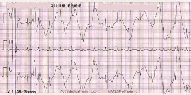
What electrodes do leads I and II have in common?
- Lead I is a dipole with the negative electrode on the right arm and the positive electrode on the left arm.
- Lead II is a dipole with the negative lead on the right arm and the positive electrode on the left leg.
Lead I and lead II share the right arm electrode! That is the electrode that is causing this problem.
Wandering Baseline Artifact
Baseline wander appears on the ECG tracing as slowly drifting up and down instead of remaining flat. It is often caused by patient movement and may follow the patient’s respiratory pattern, but it can also be caused by loose or dry electrodes.
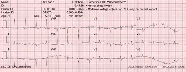
See the above ECG tracing with significant baseline wander in the precordial Leads V4-V6 which likely corresponds to the patient’s respiration.
It may be helpful to have the patient hold their breath while capturing a 12-lead ECG but be aware that this can also alter the patient’s heart rate. There are times when your patient is acutely short of breath, and it’s simply impossible to capture a 12-lead ECG with excellent data quality.
Muscle Tremor and Motion Artifact
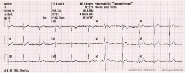
Muscle tremor (or tension) artifact is a type of motion artifact that appears “fuzzy” or “choppy”, often occurring when your patient is shivering, anxious or exerting muscle tension such as propping themselves upright. It can also be caused by neurologic conditions such as Parkinson’s disease.
The example below was obtained from a young, healthy firefighter during routine training. It was cold in the fire station, and he was shivering.
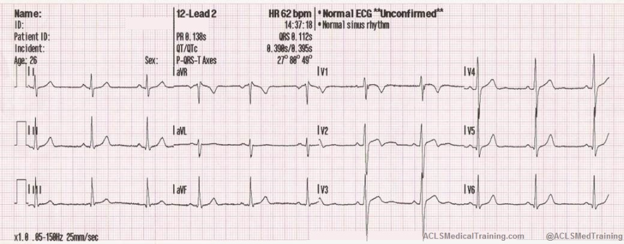
The next example was taken after a large towel was placed over the firefighter. A simple intervention made quite a difference, didn’t it? Look to address the causes of the “shaky” ECG tracing, whether with a warm blanket, relaxation techniques, or by supporting the limbs to remove muscle tension.
Electromagnetic Interference (EMI or 60 Hz Artifact)
Electromagnetic interference (EMI) artifact usually results from electrical equipment and their lines proximate to the patient, as well as cell phones. This is also known as 60 cycle interference (or 60 Hz pickup) and appears as rapid, “caterpillar-like” oscillation patterns on the ECG.

Creative Commons: ECGGuru.com
To help minimize 60 cycle interference, you may need to remove or relocate a power cord to another outlet, turn off light switches, or even remove the patient’s cell phone while conducting a 12-lead ECG. If removing power cables, ensure that they are not connected to a device performing a critical or necessary function which cannot be interrupted (e.g. diagnostic or life-support devices)! To help minimize 60 cycle interference you should also ensure the “notch filter” (60Hz) is engaged to minimize non-cardiac electrical background noise.
CPR Compression Artifact
CPR compression artifact occurs when chest compressions induce an ECG artifact that is registered on the ECG or the defibrillation pads attached to the patient. They can appear as rhythmic, high-amplitude waves that can mimic an arrhythmia such as ventricular fibrillation or ventricular tachycardia.
This ECG was automatically recorded during a cardiac arrest.
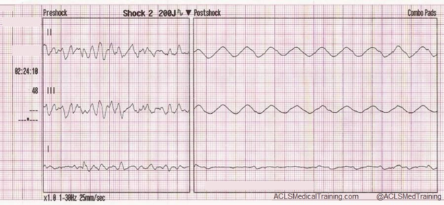
The wavy line after the shock is CPR artifact. Using the small block method (1500/13=115) we can determine that the compression rate was about 115/min, which is perfect!
CPR artifact makes it difficult to determine the underlying rhythm; however, if you’re performing CPR at a 30:2 compression to ventilation ratio, you can see the underlying rhythm during ventilations or during the pause for a rhythm check every 2 minutes.
Neuromodulation and Implantable Device Artifact
Neuromodulation devices, such as spinal cord stimulators and deep brain stimulators, can cause significant ECG distortion that mimic dangerous morphology, leading to unnecessary treatment. This type of artifact is increasing in prevalence as implantable neurostimulators become more common.
These devices are used to treat a variety of symptoms, including tremors, seizures, chronic pain, GI/GU issues, visual impairment, and hypertension.
Neuromodulation device artifact often manifests as sharp spikes which can mimic QRS complexes or pacemaker activity.
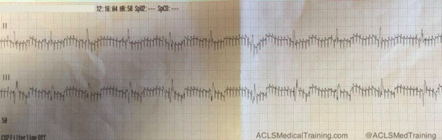
If you see an artifact that looks like this, you should ask the patient if he or she has any implantable medical devices. Some devices can be temporarily turned off with a magnet, but you should consult with the prescribing physician.
Transcutaneous Pacing Artifact (Echo Distortion or False Capture)
This type of artifact is associated with transcutaneous pacing (TCP). Echo distortion causes a pseudo-QRS complex after the pacing spike, which is sometimes referred to as “false capture.”
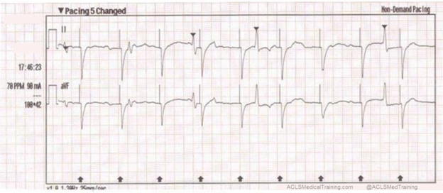
Following the pacer “spike” during TCP, the ECG monitor intentionally “blanks out” for 40 to 80 milliseconds while the powerful electrical current is transmitted through the pacer pads and chest wall to the heart. It does this so that the massive electrical current from the pacer is not registered as artifact on the ECG monitor.
After “blanking out”, the ECG then begins registering the QRS complex that is induced by the pacing stimulus. Despite this ECG design for TCP modes, the monitor still sometimes registers a portion of the massive electrical pacing current as it returns to baseline which registers as a pseudo-QRS complex on the ECG. This is problematic if the pacer did not produce a QRS, but the artifact makes it to appear as if one did.
To identify this false capture, verify whether the pacer spike is followed by a wide QRS with a T-wave, and whether a palpable pulse is felt following each QRS complex. If it is found to be artifact, relocate the ECG electrodes farther away from the pacing pads and adjust the pacing current until capture is seen for every pacing spike.
Arterial Pulse Tapping Artifact
This unusual artifact is caused by a limb electrode that picks up mechanical pulsation from placement near the radial or posterior tibial arteries. The typical lead pattern changes seen in this artifact are ST changes with large, bizarre T-waves on all but one lead in the ECG. These types of changes can be mistaken as repolarization abnormalities, since the ST changes can mimic findings of ischemia on the ECG with 1:1 rhythmic pattern following each P and QRS wave.
The phenomenon was first reported in 2005 by Özhan et al. as a “bizarre electrocardiogram” thought to be associated with abnormal left ventricular motion.
Subsequent work by Aslanger solved the issue in favor of arterial pulse tapping (which explains why the artifact occurs synchronously with the cardiac cycle on the ECG.)
Consider these two ECGs which were recorded from the same patient less than 1 minute apart. The first ECG shows simple motion artifact in leads I, III, and aVL.
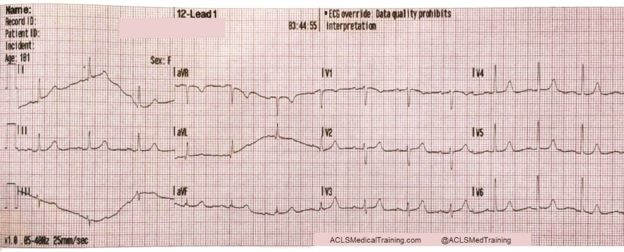
Courtesy of Frank Intessimoni (@njmedic3228)
The second ECG shows large, bizarre T-waves that were concerning to the paramedics on the call.
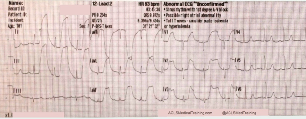
Courtesy of Frank Intessimoni (@njmedic3228)
You will note that the artifact is most pronounced in leads I, II, and aVR. Lead III appears perfectly normal. This suggests that the right arm electrode was placed over the radial artery.
But if that’s true, why is there also an artifact in other leads?
Aslanger explains:
“[O]ne may expect that the leads not connected to the electrode affected by the source of disturbance would be free of distortion, but this is not the case. When one of the limb electrodes is affected by a source of disturbance, it distorts not only the corresponding derivation but also [the others], which are all calculated by mathematical equations.”
How to Tell ECG Artifact from True Arrhythmia
When differentiating the patient’s true underlying cardiac rhythm from artifact, begin by looking for the R-R intervals of readily apparent QRS complexes. Even with a chaotic baseline, a regular R-R interval favors artifact over arrhythmia.
Next, look at your patient. If the monitor shows what looks like V-Fib or pulseless V-Tach but the patient is coherent and calm with no signs of distress, the rhythm on the screen is certainly artifact. In such cases, is the patient brushing their teeth? Shivering? Walking? Exerting themselves?
Additionally, for any true arrhythmia, the 12-lead ECG tracing should be consistently abnormal in all the leads that share the same anatomical “view” of the electrical event. Thus, if only one precordial lead (V1-V6) shows an abnormality, you can have confidence that that is likely artifact requiring at least a check of the electrode placement.
Repeat the ECG whenever:
- Artifact is suspected which inhibits measurement of the ST-segment relative to the TP segment (preventing measurement of elevation/depression).
- The tracing baseline is “fuzzy” to the point of hiding P waves.
- The EKG shows a new concerning finding that doesn’t match the patient’s stable status (V-Fib, V-Tach, marked ST elevation).
Best Practices for Preventing ECG Artifact
When considering ECG troubleshooting tips, artifact is best treated by prevention which requires proper skin preparation with consideration of an alcohol swab, non-stick barrier film, and anatomically correct electrode placement of all leads (e.g. intercostal spaces, under the breast tissue).
Additionally, artifact can be treated or prevented by looking at the patient to maximize comfort and minimize movement (e.g. blankets for shivering, relaxation for anxiety, pillows for positioning).
If artifact is seen, look for dried-out electrodes and loose or cracked lead wires. Also, ensure the leads do not cross over power cables and set notch filters on the monitor if the ECG is especially “fuzzy”.
Remember, ECG artifact management is primarily improved by training and repeated exposure troubleshooting the same artifact.
ECG Artifact Recognition in ACLS and High-Acuity Care
During cardiac emergencies, rapidly obtaining a clear ECG is paramount in diagnosing the patient’s condition and determining next steps for treatment with ACLS protocol. Recognizing artifact is an essential skill in ensuring a correct rhythm interpretation, especially in high-pressure environments where small details such as a “fuzzy” baseline or false capture can be missed. For ECG artifact, ACLS confidence and clarity allows structured thinking for the clinician when the stakes are highest for patient outcomes.
Continuing Education and Skill Reinforcement
Artifact recognition, while challenging, is enhanced via formal ECG interpretation training with repetitions in simulated scenarios. ECG interpretation skills are essential in carrying out swift and efficient BLS and ACLS protocols which contribute to positive patient outcomes. To further improve your ECG interpretation skills and prepare for critical cardiac emergencies, consider enrolling in our ACLS Certification Training course or our BLS Certification Training course . Our proven curriculum will equip you with the guidance and skills to confidently handle any cardiac emergencies that you encounter in your practice. Formalize your knowledge and skills by enrolling today !
References
Aslanger E, Yalin K. Electromechanical association: a subtle electrocardiogram artifact. Journal of Electrocardiology. 2012;45(1):15-17. doi:10.1016/j.jelectrocard.2010.12.162.
Aslanger E, Bjerregaard P. Mystery of “bizarre electrocardiogram” solved. Journal of Electrocardiology. 2011;44(6):810-811. doi:10.1016/j.jelectrocard.2011.04.001.
Goldberger AL, Burns SM. ECG tutorial: Miscellaneous diagnoses. In: Post TW, ed. UpToDate. UpToDate; 2026. Accessed February 18, 2026.
Pérez-Riera AR, Barbosa-Barros R, Daminello-Raimundo R, de Abreu LC. Main artifacts in electrocardiography. Ann Noninvasive Electrocardiol. 2018;23(2):e12494. doi:10.1111/anec.12494
Vreugdenhil S, de Heus RA, Post B, van der Lely AJ, Vecht J, van Vugt JPP. Neurostimulator-induced ECG artefacts: A systematic analysis. Clin Neurol Neurosurg. 2021;203:106557. doi:10.1016/j.clineuro.2021.106557
